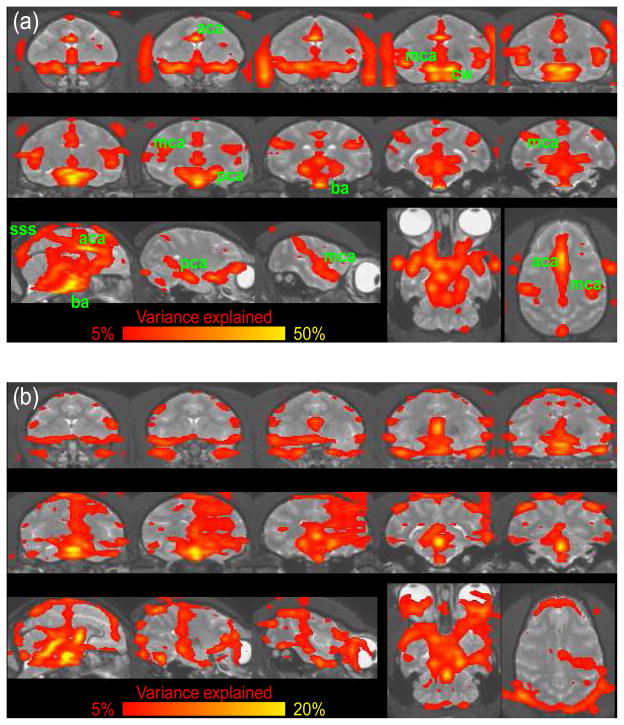
Fig. 1.
Cardiac (a) and Respiratory Artifact (b) from one macaque monkey. Data were averaged over 8 runs of 400 volumes each. Regions with significant cyclic artifacts are overlaid on the individual T1-weighted structural image. The colors correspond to the percent variance explained by the harmonic regression. Note the different scales of the color bar for the cardiac and respiratory artifacts. (a) Regions with significant artifacts can be found around major vessels. The basilar artery (ba), anterior (aca), medial (mca) and posterior (pca) cerebral arteries can clearly be distinguished. Veins seem to cause less artifacts but the superior sagital sinus (sss) is clearly visible. Arteries outside the brain such as the opthalmic artery are also visible. (b) Part of the respiratory artifacts are caused by breathing related apparent motion.

