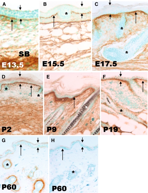
Fig. 1.
Detection of periostin protein deposition in development by immunohistochemistry. At E13.5 (a), periostin labeling in dermal-epidermal junction (long arrow) was faint and uneven, in contrast the subcutaneous tissue (SB) was strongly labeled. As development advanced (b, c, d, e), periostin immunoreactivity within the DEJ increased, and was maintained within the dermis. The outline of HF (*) remained negative till P9 (e). Compared with P9, the labeling within the DEJ was reduced at P19 when HF(*) was faintly labeled. In adults (P60 days), periostin was faintly detectable within the DEJ, whilst the ECM surrounding hair follicles was strongly labeled (g). As a negative control, the periostin null skin (h) showed no labeling either in the DEJ or surrounding the HFs. All images were taken at same magnification. Scale bar = 200 µm

