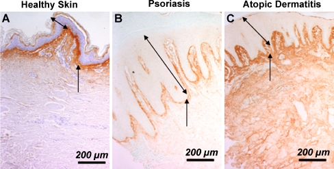
Fig. 5.
Immunohistochemical detection of periostin in human skin samples. Periostin was detected in healthy skin (a), skin of psoriasis (b) and skin of atopic dermatitis (c) skin biopsy samples. In healthy skin (a) and skin of psoriasis (b) patients, the periostin protein is predominantly deposited along the DEJ. However, in skin from atopic dermatitis (c) patients, the deposition is enhanced and more widely distributed with whole the dermis labeled positive. Note the epidermal thickness is significantly increased in both types of skin disease compared to healthy skin. The two-headed arrows denote the epidermal thickness and the arrows point to the DEJ. Bar = 200 µm

