
Figure 3.
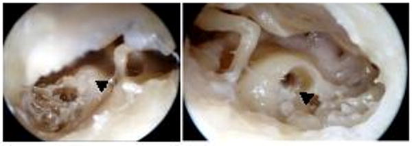
Photographs illustrating the cement-ossified cochlea (A) posterior tympanotomy (left ear) showing Simplex B cement filling the cochleostomy (arrow). (B) right cochleostomy after drill out of cement-ossification. A rim of residual cement is still present (arrow).