Fig. 3.
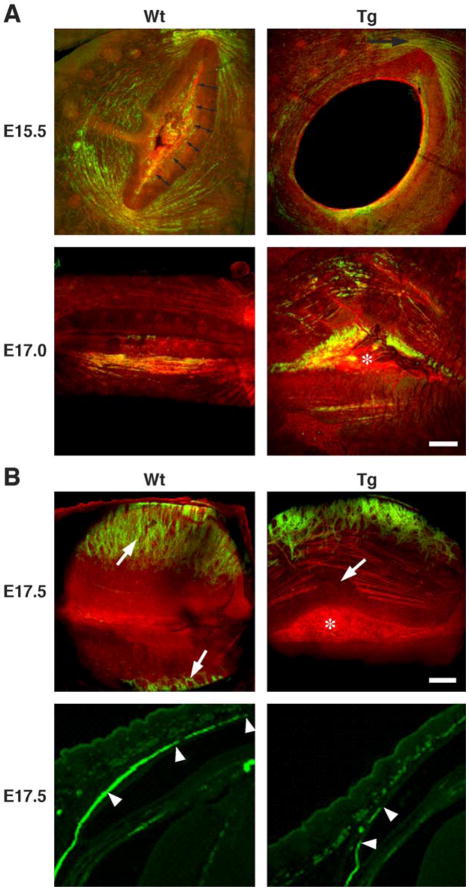
Development of eyelid orbicularis oculi, and levator palpebrae and tarsal muscles. (A) Confocal three-dimensional maximum intensity projections of the outer layers (epidermal side) of the developing orbicularis oculi from wild type (Wt) and Kera-Bgn (Tg) mice at E15.5 and E17.0. Lids are stained by anti-α-SMA staining (green) and phalloidin (red). Developing orbicularis oculi muscle shows continuous anti-α-SMA staining circumferentially around the eyelids and presumptive tarsal plate (double headed arrows) in wild type (Wt). At the same developmental stage, Kera-Bgn mice showed muscle development limited to the medial and lateral (top right panel, arrow) aspects of the eyelids. This developmental stage was approximately 12 h behind that detected for the wild-type (Wt) lid. The presumptive tarsal plate never appeared to become compacted, and the area is covered by remnant epidermal epithelium (asterisk). (B) Confocal three-dimensional maximum intensity projections of the outer layers (top panels, palpebral side) and immunofluorescent staining (bottom panels) of the developing eyelid from wild type (Wt) and Kera-Bgn (Tg) mice at E17.5. In wild-type mice, the tarsal muscle bundles run vertically toward the lid tip (top left panel, arrows; bottom left panel arrowheads). In Kera-Bgn (Tg) mice, the lid margins never fuse (top right panel, asterisk) with breaks in the developing orbicularis muscle fibers (top right panel, arrow). Development of the tarsal muscles is also delayed and fragmented (right bottom panel, arrowheads).

