Fig. 4.
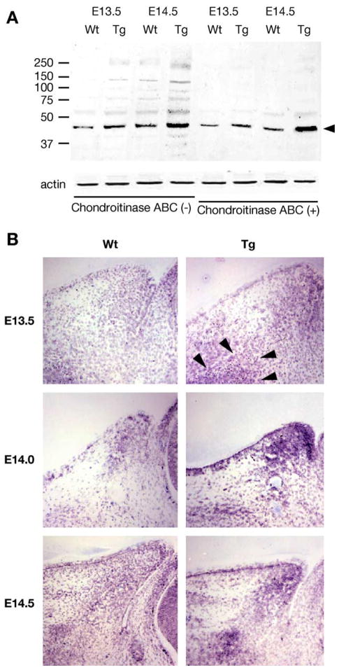
Expression of biglycan determined by Western blot and in situ hybridization. (A) Protein extracts of eyelids from E13.5 and E14.5 wild type (Wt) and Kera-Bgn (Tg) mice were treated with and without chondroitinase ABC (0.5 unit/ml) prior to Western blot analysis with rabbit anti-biglycan antibodies. Actin determined by Western analysis was used to normalize the amounts of proteins applied to the gels. The biglycan existed as a heterogeneous CS/DS proteoglycan with molecular weights ranging from 46 kDa to greater than 250 kDa. The high molecular proteins reacted by the antibodies were digested by the enzyme and migrate as the 46 kDa biglycan core protein. (B) In situ hybridization reveals that biglycan mRNA was expressed by migrating mesenchymal cells of eyelid from wild-type (Wt) mice. In Kera-Bgn (Tg) mice, migrating mesenchymal cells (arrowheads) expressing biglycan accumulate under the epithelium adjacent to the palpebral side of conjunctiva at the tip of the lid from E13.5 through E14.5, with an interrupted pattern throughout the eyelid stroma as compared to wild-type (Wt) mice at E14.5.

