
Figure 2. Mycobacterial killing by Ubiquitinated Peptides.
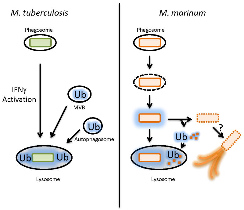
Left, Model for killing of M. tuberculosis by ubiquitin peptides by macrophages. In IFNγ activated macrophages, the M. tuberculosis phagosome is thought to fuse with ubiquitin (Ub) -containing vesicles including MVBs (multivesicular bodies generated by the ESCRT complex) and LC-3 positive (red line) autophagosomes, and the ubiquitin coated bacteria are targeted to the lysosome. Right, M. marinum differs from M. tuberculosis in that a population of M. marinum escape the phagosome. Cytosolic M. marinum are coated with ubiquitin in an autophagy-independent manner. The coated M. marinum have two possible fates. First, they can be targeted to the lysosome. Second, the bacteria can shed the ubiquitin coated cell wall (which itself is sent to the lysosome). It is possible that this population then forms actin tails, the actin tail population is not ubiquitinated.