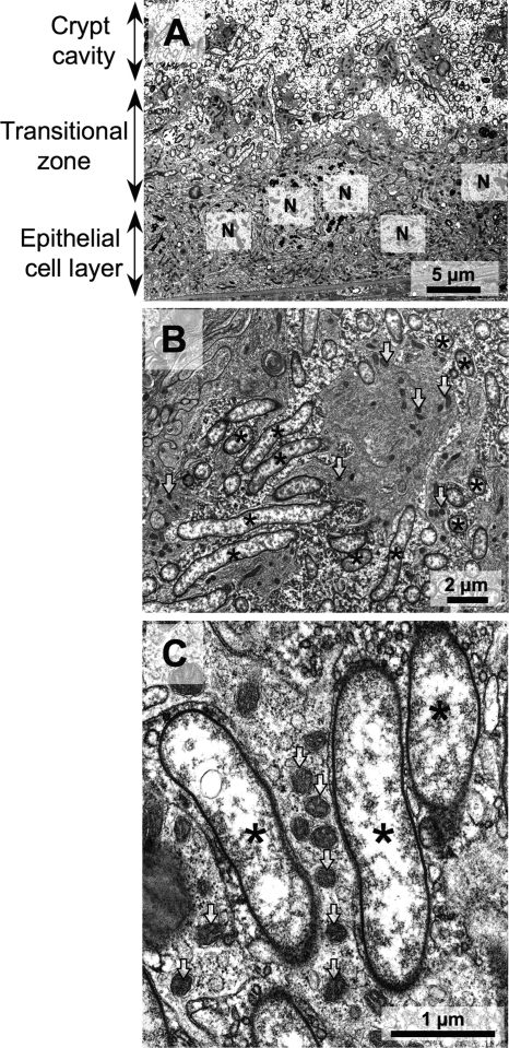
FIG. 5.
Transmission electron microscopy of the midgut crypts in adult females of C. ocellatus. (A) Image of the interface between the crypt cavity and the epithelial cell layer; (B) enlarged image of the transitional zone; (C) enlarged image of the symbiont cells indicating an extracellular, rather than endocellular location. Asterisks and arrows indicate the symbiont cells and mitochondria, respectively. N, nucleus of the epithelial cell of the midgut crypt.

