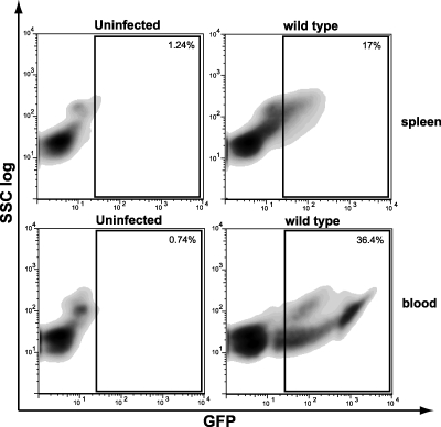
FIG. 5.
In vivo and ex vivo infection by L. monocytogenes EGD-cGFP: flow cytometry data for uninfected and infected spleens (in vivo) and blood (ex vivo). BALB/c mice were infected with 105 CFU of L. monocytogenes wild-type strain EGD intravenously for 3 days. Blood was collected from BALB/c mice and infected with 109 wild-type L. monocytogenes CFU/ml of blood. Spleen and blood cells were collected and analyzed by FACS. At least 20,000 cells were analyzed for each sample, and the data are representative of the results of three experiments. Dot plots and gates were obtained using FlowJo software.

