FIG. 1.
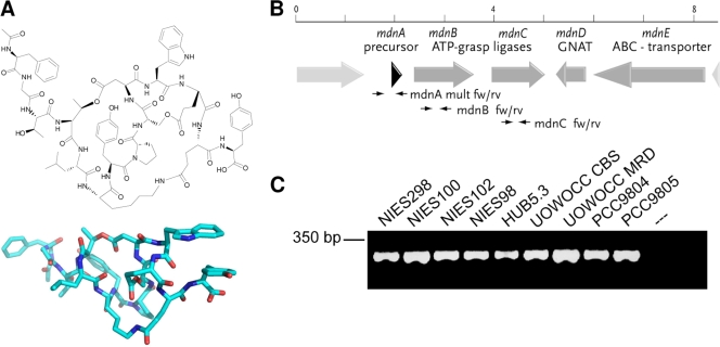
(A) Representative structure of microviridin B and three-dimensional model. (B) Schematic representation of the microviridin gene cluster of M. aeruginosa NIES298. The positions of primers for the amplification of mdnA, -B, and -C are indicated. GNAT, GCN5-related N-acetyltransferase. (C) PCR gel picture showing the amplification of mdnB from a selection of laboratory strains.

