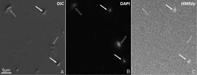
FIG. 3.
Fluorescent in situ hybridization (FISH) of cells of strain HSMV-1 using an HSMV-1-specific oligonucleotide rRNA probe (HSMVp). Cells used for FISH were magnetically concentrated by placing a magnet next to the side of the sample bottle for 30 min and then removed with a Pasteur pipette. This technique was used rather than the magnetic racetrack method in order to have many HSMV-1 cells as well as some other cells that could be used as a negative control. (A) Differential interference contrast (DIC) image of HSMV-1 cells (filled arrows) and other cells (negative control; empty arrows) from hot spring samples; (B) cells stained with 4′,6-diamidino-2-phenylindole (DAPI); (C) cells hybridized with the specific probe HSMVp.

