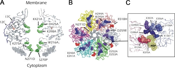
FIG. 3.
Representations of the 3D structure of TrwBΔN70 bound to ADP (PDB accession no. 1GKI), where the mutated residues are highlighted in the space-filled format. (A) Side view of two opposing monomers, showing in green the mutated residues which protrude into the ICH of the hexamer. The extent of the 12 and 17 C-terminal residues of the protein is indicated with arrows (12C and 17C, respectively). Mutated AAD residues are also indicated (space filled in gray). (B) TrwBΔN70 hexamer seen from the cytoplasmic side, with the selected residues from the AAD domain shown in the space-filled format. Residue R318, located in the interface between monomers, is also highlighted (in red). The region included in the quadrangle is amplified in panel C for better resolution of the mutated residues in the proximity of the nucleotide molecule.

