
FIG. 3.
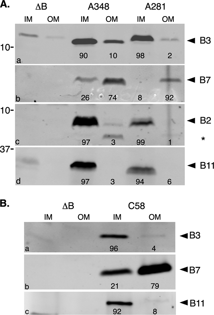
Membrane localization of VirB3. Subcellular locations of VirB3 expressed from the Ti plasmids pTiA6NC (A), pTiBo542 (A), and pTiC58 (B) were determined by Western blot assays. Inner (IM) and outer (OM) membranes were separated by sucrose density gradient centrifugation, and the presence of VirB proteins was monitored by Western blot assays. Proteins were separated in SDS-12.5% PA-Tricine gels and transferred to nitrocellulose filters. (a) The filter was probed with anti-VirB3 antibodies. For control experiments, the filter in panel a was probed with anti-VirB11 antibodies (d), and a duplicate filter was probed sequentially with anti-VirB7 (b) and anti-VirB2 (c) antibodies. The residual VirB7-specific band in panel c is marked with an asterisk. Bands were quantified by measuring pixel volume, using Li-Cor Odyssey software. The number below each lane indicates the percentage of the protein present in that fraction. ΔB, A348ΔB; A348, octopine strain with pTiA6NC; A281, supervirulent strain with pTiBo542; C58, nopaline strain with pTiC58.