FIG. 2.
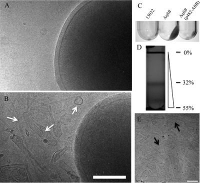
Identification of membrane fragments in the culture medium of the ΔaftB mutant. (A and B) Cryo-electron microscopy images of C. glutamicum 13032 and ΔaftB cells grown on BHI medium, respectively. Culture supernatant from the wild type, the ΔaftB mutant, or the complemented strain was ultracentrifuged as described in Materials and Methods. An important pellet is clearly visible for the mutant strain but not for the wild type or the complemented strain (C). Membrane fragments released by the ΔaftB strain were then purified on a sucrose gradient (D) and were observed directly by cryo-electron microscopy (E). Fragments of different sizes and forms are indicated by arrows. The percentages of sucrose (wt/wt) at specific positions are indicated next to the gradient. Bars, 200 nm.

