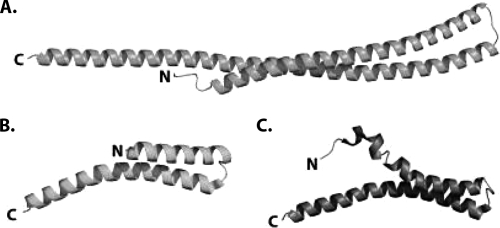
FIG. 6.
Comparison of the structures of CT670 and the type III needle proteins MxiH and PrgI. (A) CT670 in the orientation shown in Fig. 1A. (B) MxiH (PDB code CA5) from S. flexneri. (C) PrgI (PDB code 2JOW) from S. enterica serovar Typhimurium. The structures are represented by ribbons, and the N and C termini are labeled.

