FIG. 3.
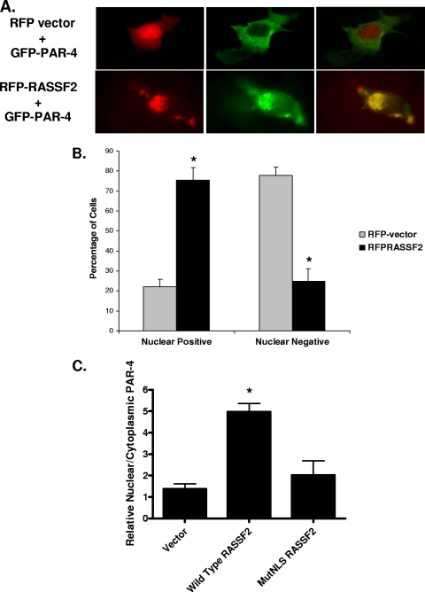
RASSF2 promotes the nuclear localization of PAR-4. (A) COS-7 cells were cotransfected with RFP vector or RFP-RASSF2 and GFP-PAR-4, and images were captured 24 h later using a fluorescence microscope as described in Materials and Methods. In the absence of RASSF2, PAR-4 localized to the plasma membrane and cytoplasm, but in the presence of RASSF2, PAR-4 colocalized with RASSF2 in the nucleus, as evidenced by the yellow color in the merged image. Magnification (all images), ×100. (B) Quantification of PAR-4 nuclear localization in the presence of RASSF2. Fifty randomly selected cells expressing both RFP-RASSF2 or RFP-vector and GFP-PAR-4 were scored for the presence of nuclear PAR-4. The bars show the means of triplicate experiments, and standard deviations are indicated. *, statistically significantly different (P < 0.01) from vector-transfected cells. (C) PC-3 human prostate tumor cells were transfected with RASSF2 or a deletion mutant of RASSF2 lacking at least one of the NLSs (MutNLS) and fractionated before Western blotting for endogenous PAR-4. The blots were quantified and used to generate a ratio of nuclear versus cytoplasmic PAR-4. The mutant of RASSF2 was impaired for inducing the nuclear localization of endogenous PAR-4. *, statistically different (P < 0.01) from vector-transfected cells.

