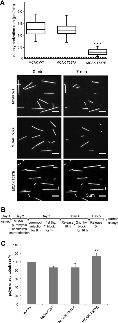
FIG. 3.
Phosphomimetic MCAK T537E attenuates its microtubule-destabilizing activity in vitro as well as in vivo. (A) In vitro depolymerization assays. (Upper panel) Box plots depicting the distribution of depolymerization rates of individual microtubules observed upon addition of MCAK WT (n = 45), MCAK T537A (n = 44), or MCAK T537E (n = 59). The box depicts the range between the 1st and 3rd quartiles (i.e., central 50% of the distribution), with the central line showing the position of the median. ***, P < 0.001. The dashed line shows the spontaneous depolymerization rate for the stabilized microtubules used in these assays (0.03 μm/min). (Lower panel) Represen tative epifluorescence images of immobilized microtubules either prior to or 7 min after addition of MCAK WT, T537A, or T537E. Bar, 5 μm. (B) Working schedule. (C) Quantification of polymerized tubulin content in vivo. HeLa cells were transfected with Flag-tagged MCAK WT, its mutants, or empty vector in an endogenous MCAK-depleted background. Transfected cells were then synchronized to the G1/S boundary and released for 10 h. Cellular polymerized tubulin contents were analyzed by flow cytometry after cells were extracted, fixed, and stained for tubulin. The amount of polymerized tubulin from Flag vector plasmid-transfected HeLa cells was assigned as 100%. The results are presented as means ± standard deviations (SD) (n = 3). **, P < 0.01 for comparison to MCAK WT.

