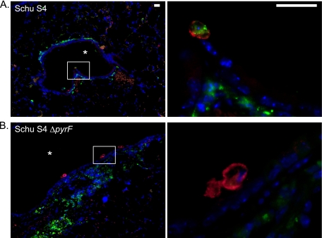
FIG. 11.
Immunofluorescence microscopy of lung sections from mice infected with F. tularensis Schu S4 or Schu S4 ΔpyrF. Mice were infected i.t. as described in the legend to Fig. 7. On day 4 postinfection, lungs were harvested from mice infected with Schu S4 (A) or Schu S4 ΔpyrF (B), were fixed in formalin, and were subsequently embedded in paraffin. Deparaffinized lung sections were probed with rat anti-mouse F4/80 to detect macrophages and with rabbit anti-F. tularensis. The secondary antibodies used were Alexa Fluor 555 goat anti-rat immunoglobulin and Alexa Fluor 488 donkey anti-rabbit immunoglobulin. DNA was stained with DAPI. For both panels, the color images represent a three-color merge (red, F4/80; green, F. tularensis; and blue, DAPI). The first color image represents low-power magnification, and the second image is a high-power magnification of the boxed section. In the low-power images, the asterisk designates a major airway. The scale bars in the upper right hand corners of the images in panel A represent a length of 20 μm.

