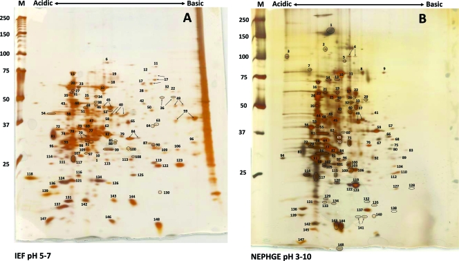
FIG. 1.
Two-dimensional gel electrophoresis of T. pallidum proteins. T. pallidum lysates were separated by IEF at pH 5 to 7 (A) or NEPHGE at pH 3.5 to 10 (B) in the first dimension, followed by 8 to 20% SDS-PAGE in the second dimension. Gels were subsequently silver stained for protein visualization. Acidic and basic ends are denoted, and relative molecular mass markers (in kilodaltons) are indicated to the left of each gel. A T. pallidum lysate, resolved in the second dimension only, is shown at the right side of the IEF pH 5-to-7 gel (A). The identities of the numbered spots are presented in Table 1. Arrows indicate spots that were submitted separately for MALDI-TOF MS but returned the same identity. Circles demarcate some closely spaced spots to indicate more clearly which spots are labeled.

