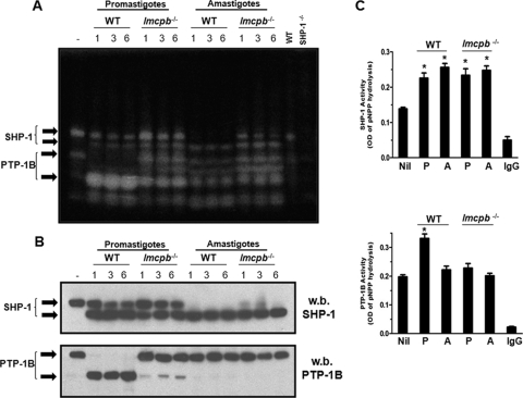
FIG. 3.
Role of LmCPb in PTP-1B activation. (A) B10R Mφs were infected with L. mexicana promastigotes and amastigotes of WT and lmcpb−/− parasites for the specified time course (0 to 6 h, 20:1 ratio), and cell lysates (30 μg) were subjected to an in-gel PTP assay. The rightmost two lanes contain lysates (30 μg) from WT and SHP-1−/− Mφs to confirm the band corresponding to SHP-1. (B) Samples in panel A were run on gels by SDS-PAGE and blotted for SHP-1 (top panel) and PTP-1B (bottom panel). w.b., Western blotted. (C) B10R Mφs were infected with L. mexicana promastigotes (P) and amastigotes (A) of WT and lmcpb−/− parasites (3 h, 20:1 ratio) followed by lysis and immunoprecipitation of SHP-1 or PTP-1B. The SHP-1 and PTP-1B IPs were subjected to a pNPP phosphatase assay (top and bottom graphs, respectively). The values are means plus standard errors of the means (SEMs) (error bars). Values that were significantly different (P < 0.05, ANOVA test) are indicated by an asterisk. Rabbit anti-rat IgG was used as a negative control for the IPs. All results are representative of at least three independent experiments.

