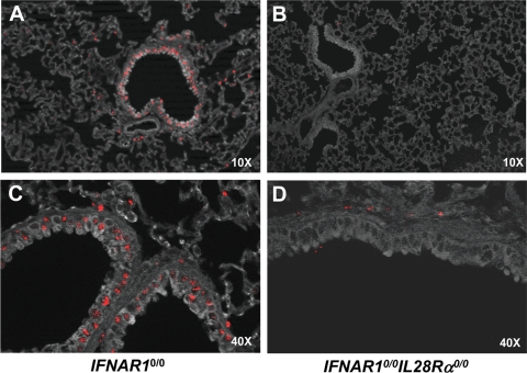
FIG. 6.
Lung epithelial cells express functional IFN-λ receptor complexes. IFNAR10/0 (A, C) and IFNAR10/0IL28Rα0/0 (B, D) mice were intranasally infected with 5 × 10⁴ PFU of SC35M-ΔNS1, a potent inducer of type I and type III IFN (26). At 20 h postinfection, the lungs were removed and stained for Mx1 by immunohistofluorescence. (A, B) Low magnification overview. Mx1-positive cells (stained nuclei) are mainly clustered around bronchioles in IFNAR10/0 mice but mostly absent in IFNAR10/0IL28Rα0/0 mice. (B, D) High magnification of bronchioles and surrounding tissue. Epithelial cells were prominently stained for Mx1 in IFNAR10/0 but not in IFNAR10/0IL28Rα0/0 mice.

