
FIG. 2.
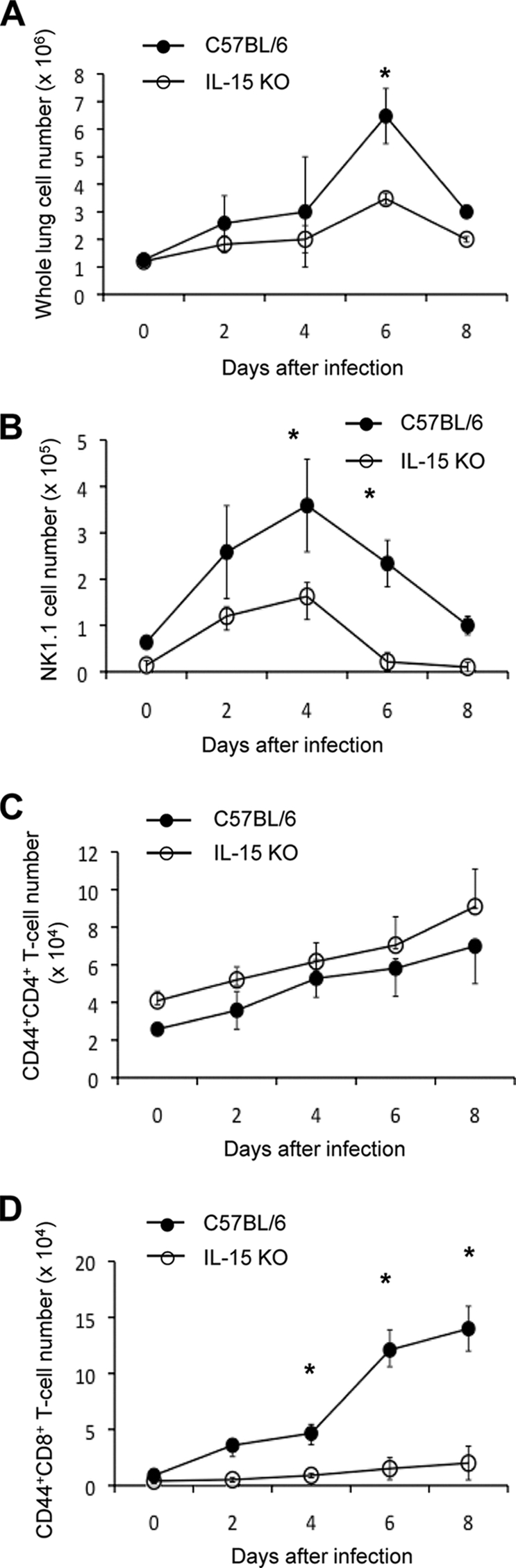
Cell population infiltrating the lungs of IL-15 KO mice after infection with influenza virus A/FM/1/47. The lung cells were isolated on days 2, 4, 6, and 8 after infection with 500 PFU of influenza virus A/FM/1/47, and the absolute number of lung cells was counted (A). The cells were stained with anti-NK1.1, CD4, CD8, or CD44 MAb and subjected to flow cytometric analysis. The absolute numbers of NK1.1+ cells (B), CD44+ CD4+ T cells (C), and CD44+ CD8+ T cells (D) were counted by multiplying the percentage of the cells by the absolute number of lung cells. Each group consisted of 5 mice. Statistically significant differences (P < 0.05) between C57BL/6 and IL-15 KO mice are indicated with n asterisk. Each point shows the mean ± standard deviation (SD) (error bar). Data are representative of at least three independent experiments.