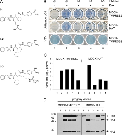
FIG. 6.
Suppression of HA cleavage and influenza virus spread by protease inhibitors. (A) Structural formulas of peptide mimetic inhibitors I-1, I-2, and I-3. (B) MDCK-TMPRSS2 and MDCK-HAT cells were infected with A/Hamburg/09 (H1N1) at an MOI of 0.02 and VSV at an MOI of 0.001, respectively, and incubated with or without doxycycline (Dox) in the absence (Ø) or presence of inhibitor I-1, I-2, or I-3 at a final concentration of 30 μM at 37°C. At 24 h p.i. the cells were immunostained with influenza virus NP- and VSV-specific antibodies, respectively. (C) MDCK-TMPRSS2 and MDCK-HAT cells were infected with A/Hamburg/09 at an MOI of 0.01 and incubated in the absence or presence of doxycycline for 24 h (lanes 1 and 2, see panel B) in the absence or presence of inhibitor I-1 (lane 3), I-2 (lane 4), or I-3 (lane 5). Virus titers were determined by plaque assay at 24 h p.i. Note that the bars representing virus titers in cells without induction of TMPRSS2 and HAT expression (−Dox), respectively, cannot be seen in the figure since the titers were below the limit of determination (102 PFU/ml). The results are the mean values of two independent experiments. (D) Virus-containing cell supernatants shown in Fig. 6C were concentrated by ultracentrifugation and then subjected to SDS-PAGE under reducing conditions and Western blot analysis with HA-specific antibodies.

