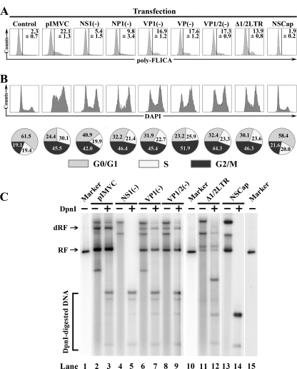
FIG. 3.
Replication of the MVC genome activates caspases, and the MVC genome per se arrests cell cycle at the G2/M phase. WRD cells were transfected with plasmids as shown. (A) At 48 h posttransfection, transfected cells were costained with anti-NS1, except for pIMVCNS1(−)-transfected cells, which were costained with anti-NP1 and poly-FLICA peptide. The anti-NS1-positive or anti-NP1-positive population was selectively gated and plotted as cell counts and FLICA signal. The percentage of FLICA-positive cells is shown as an average with a standard deviation generated from three independent experiments. (B) At 48 h posttransfection, transfected cells were costained with anti-NS1, except for pIMVCNS1(−)-transfected cells, which were costained with anti-NP1 and DAPI. Anti-NS1- or anti-NP1-stained cells were selectively gated and plotted as cell counts and DAPI signal. The percentage of each cell cycle phase was quantified and is shown as a pie graph at the bottom of the panel. A representative of two independent experiments is shown. (C) Southern blotting analysis of transfected WRD cells. At 48 h posttransfection, transfected cells were harvested and Hirt DNA was prepared. Hirt DNA was then digested with DpnI. The blot was probed with the NSCap probe as previously described (60). Detected bands are indicated with their respective designations to the left. Lanes 1, 10, and 15 are size markers of 5.15 kb. RF, replicative form; dRF, double replicative form.

