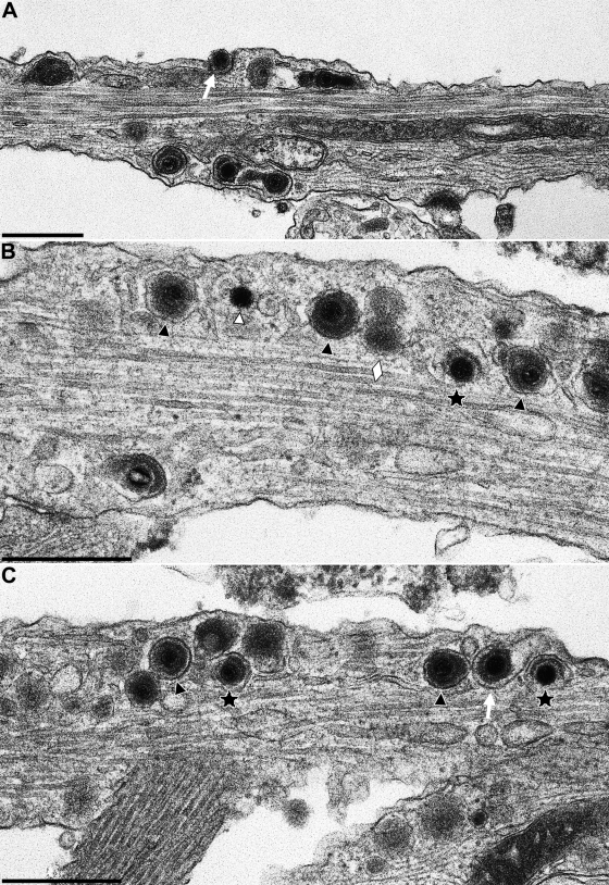
FIG. 5.
Nucleocapsids and enveloped virions in axons of PrV-infected in vitro-cultivated rat neurons. Neurons were explanted, infected with PrV-Ka, and analyzed 18 h after infection. (A to C) Three representative sections are shown. Stars indicate partially enveloped particles, white triangle highlights a nucleocapsid, black triangles show enveloped virions, and lozenges denote neurovesicles. The white arrow in panel A marks a virion in egress. Bars: 500 nm.

