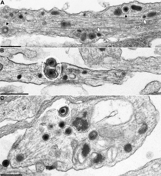
FIG. 7.
In vitro-cultivated PrV-gB− infected neuron. Neurons were explanted, infected with phenotypically complemented PrV with gB deleted that is unable to reenter cells after a first round of replication, and analyzed 18 h after infection. (A) Representative section through an axon. Black triangles indicate enveloped virions, the white triangle denotes a naked capsid, white lozenges highlight neurovesicles. (B and C) Viral egress by exocytosis along the axon (B) and at the growth cone (C). Egress stages are circled. Bars: 500 nm.

