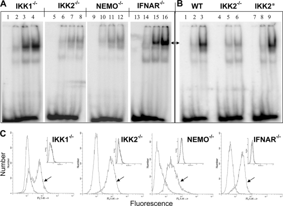
FIG. 2.
Status of canonical and noncanonical pathways of NF-κB activation upon JEV infection. (A) EMSA was performed with uninfected and JEV-infected IKK1−/−, IKK2−/−, NEMO−/−, and IFNAR−/− MEFs as indicated on top. Lanes 1, 5, 9, and 13 represent the oligomer alone; lanes 2, 6, 10, and 14 represent uninfected MEFs; lanes 3, 7, 11, and 15 represent cells infected for 10 h; and lanes 4, 8, 12, and 16 represent cells infected for 14 h. (B) EMSA was performed with wild-type (WT) and IKK2−/− MEFs and IKK2−/− MEFs that were reconstituted with the wild-type IKK2 transgene (IKK2*). Lanes 1, 4, and 7 represent oligomer alone, and lanes 2, 5, and 8 represent uninfected cells, while lanes 3, 6, and 9 represent infected cells. Arrows in panels A and B show the positions of the specific NF-κB complex. (C) As labeled, IKK1−/−, IKK2−/−, NEMO−/−, and IFNAR−/− MEFs that were infected with JEV for 12 h were stained for the expression of intracellular JEV antigen. Arrows indicate staining with primary antiflavivirus group-specific MAb followed by fluorescein isothiocyanate (FITC) anti-mouse secondary antibody, while unmarked histograms indicate staining with secondary antibody alone. The two histograms overlap in the insets, which represent staining of uninfected cells.

