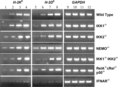
FIG. 4.
RT-PCR analysis of classical MHC-I gene (H-2Kb and H-2Db) expression in infected cells. RT-PCR analysis was carried out for 30 cycles with uninfected and JEV-infected wild-type, IKK1−/−, IKK2−/−, NEMO−/−, IKK1−/− IKK2−/−, RelA−/− cRel−/− p50−/−, and IFNAR−/− MEFs as indicated. Lanes 1, 5, and 9 represent cells that were mock infected for 36 h. Lanes 2, 6, and 10 represent results obtained at 12 h after infection. Lanes 3, 7, and 11 represent cells obtained at 24 h p.i., while lanes 4, 8, and 12 represent cells at 36 h after infection.

