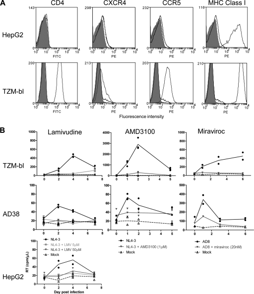
FIG. 2.
Expression and function of HIV coreceptors on hepatic cell lines. (A) Expression of CD4, CXCR4, and CCR5 was quantified using flow cytometry. Histograms of fluorescence are shown for cells alone (solid gray), isotype control (black line), and cells stained with fluorescently labeled antibodies to CD4, CXCR4, CCR5, and MHC class I (dark gray line) in HepG2 (upper row) and TZM-bl (lower row) cells. (B) Infection of AD38, HepG2, and TZM-bl cells was performed with either NL4-3 or AD8 in the absence presence of drug, or mock infection was performed. HIV reverse transcriptase (RT) in cell culture supernatant was measured. Lamivudine (at either 5 μM or 50 μM (left panels) or AMD3100 (1 μM) (middle panels) was incubated with cell lines for 24 h prior to addition of NL4-3. Maraviroc (20 nM) (right panels) was incubated with cell lines for 24 h prior to addition of AD8. Duplicates of the same experiment are shown as symbols. The line represents the mean of the duplicates.

