FIG. 3.
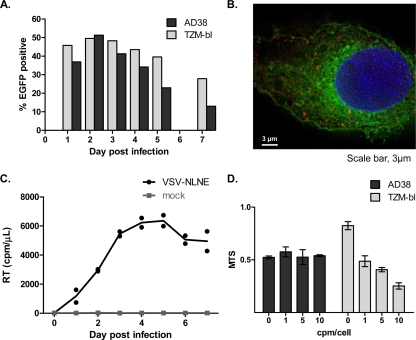
Infection of hepatic cell lines with VSV-NLNE. (A and B) AD38 and TZM-bl cells were infected with VSV-NLNE, and infection was quantified by detection of EGFP using flow cytometry (A) or fluorescence microscopy (B). An AD38 cell is shown. Hoechst staining of nucleus is blue, HBcAg staining using primary polyclonal rabbit antibody (Dako) and Texas Red-conjugated secondary goat anti-rabbit antibody (Invitrogen) is red, and EGFP expression in the HIV-infected cell is green. Magnification, ×100. (C) RT in cell culture supernatant was measured following infection of AD38 cells with VSV-NLNE or VSV-NLA1. Duplicate results from the same experiment are shown as symbols. The line represents the mean of the duplicates. Data are representative of three separate experiments. (D) MTS in AD38 cells and TZM-bl cells was measured at different multiplicities of infection (from 0 to 10 cpm/cell). The mean ± standard error (SE) for separate experiments performed in triplicate is shown.

