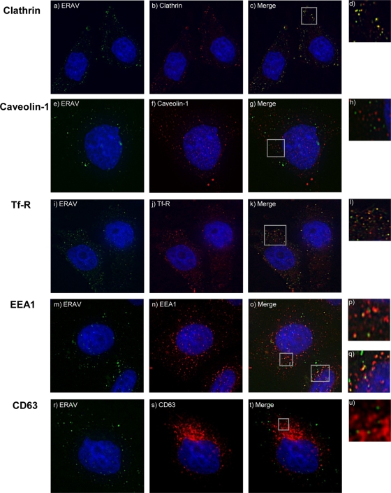
FIG. 2.
ERAV colocalization with endocytosis markers. Cells were infected with Cy2-labeled ERAV for 0 to 30 min before being visualized as described in the text. Antibodies against endocytosis markers were used in combination with secondary antibodies conjugated to Alexa-594 (Invitrogen). Cell nuclei were stained with Hoechst. Images were acquired using a DeltaVision deconvoluting microscope. (a) ERAV-Cy2 (green). (b) Clathrin (red) at 10 min pi. (c) Merge of panels a and b, showing colocalization (yellow) of ERAV and clathrin. (e to t) Cells fixed at 20 min pi with ERAV-Cy2 (green) and caveolin-1, Tfr-R, EEA1, or CD63 (red). (g) Overlay of panels e and f, showing ERAV (green) and caveolin-1 (red). (k and o) Colocalization (yellow) of ERAV with Tfr-R or EEA1, respectively. Panel k is a merge of panels i and j, and panel o is a merge of panels m and n. (t) Overlay of panels r and s, showing ERAV (green) and CD63 (red). Panels d, h, l, p, q, and u are enlargements from the Merge panels.

