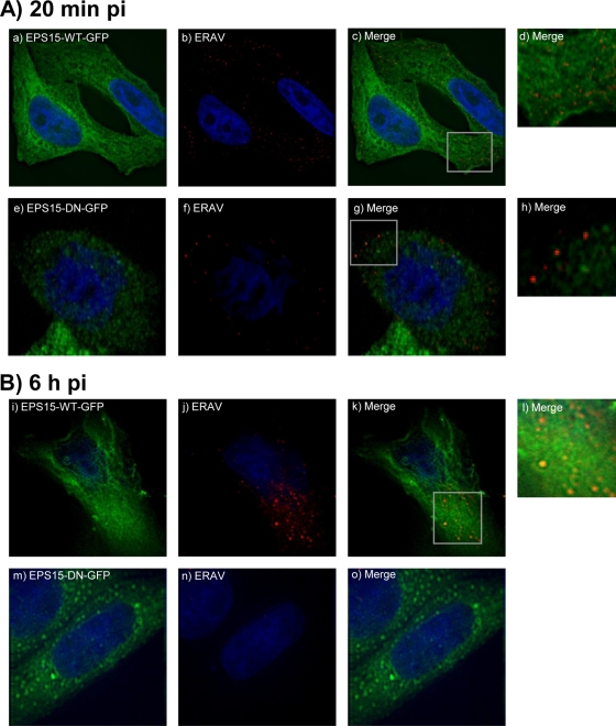
FIG. 3.
Effects of inhibition of clathrin-mediated endocytosis on ERAV entry. (A) Detection of ERAV (red) 20 min pi (b and f) of cells expressing wild-type Eps15 (Eps15-WT-GFP) (green) (a) or dominant negative Eps15 (Eps15-DN-GFP) (green) (e), which inhibits clathrin-mediated endocytosis. ERAV is located predominantly in the cytoplasm in cells expressing wild-type Eps15 (c, overlay of a and b). Panel d is an enlargement from panel c. ERAV is located predominantly at the cell surface in cells expressing dominant negative Eps15 (g, overlay of panels e and f). Panel h is an enlargement from panel g. (B) Panels i to o show cells fixed at 6 h pi. Panel k is a merge of panels i (Eps15-WT-GFP [green]) and j (ERAV [red]). The large ERAV signal in the cytoplasm is indicative of replication. Panel o is a merge of panels m (Eps15-DN-GFP [green]) and n (ERAV [red]). The lack of ERAV signal in the cytoplasm suggests lack of replication.

