FIG. 1.
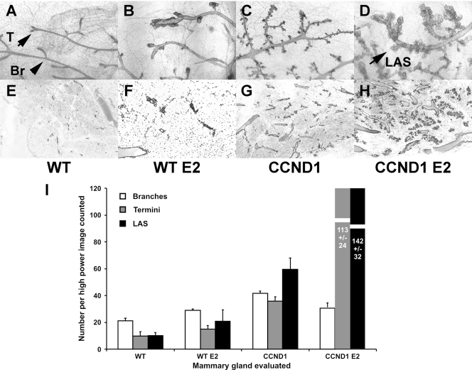
Alveolar structure numbers increase in mammary tissues constitutively expressing cyclin D1 after treatment with estrogen. Continuous release estradiol pellets were implanted subcutaneously into wild-type mice (B and F) and cyclin D1 transgenic mice (D and H) at 2 months of age as described in Materials and Methods. Control mice (A and E) were subjected to sham operations at the same time as control cyclin D1 transgenic mice (C and G). Mammary glands were harvested 2 months later for analysis using whole-mount preparations (A to D) and for routine hematoxylin and eosin staining histology (E to H). (A to D) Whole-mount photomicrographs (×10). (A) Wild-type mice (WT); (B) estradiol (E2)-treated WT mice; (C) transgenic MMTV-CCND1 mice (CCND1); (D) MMTV-CCND1 mice treated with estradiol (CCND1 E2). (E to H) Histologic photomicrographs (×40). (E) Wild-type mice (WT); (F) estradiol (E2)-treated WT mice; (G) transgenic MMTV-CCND1 mice (CCND1); (H) MMTV-CCND1 mice treated with estradiol (CCND1 E2). (I) Morphometric counts. We harvested whole-mount preparations for eight mice in each of the four groups (wild-type sham, wild-type E2, CCND1 sham, and CCND1 E2). Morphometric analyses were then performed by counting structures in three photomicrographs of each of the 32 whole-mount preparations. Numbers of branches, termini, and lobuloalveolar structures (LAS) were counted on coded photographs and totaled for each image. The means and standard errors for the numbers of branches, termini, and alveolar structures per image are shown.

