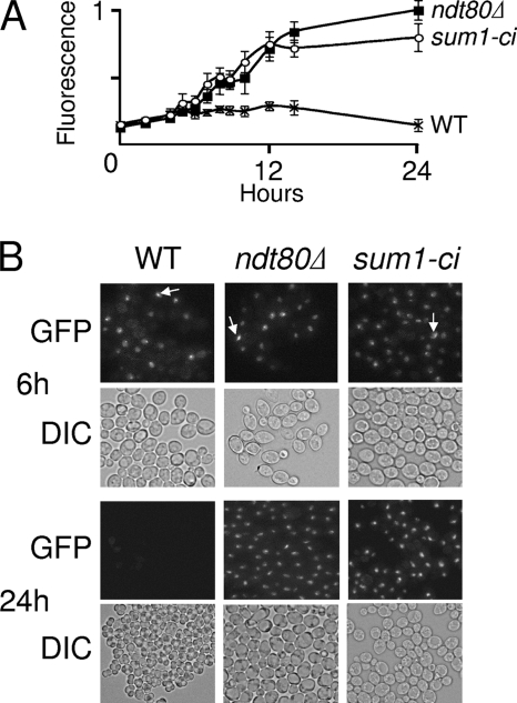
FIG. 4.
sum1-ci and ndt80Δ phenotypes are similar. (A) ZIP1-GFP (WT), ndt80Δ ZIP-GFP (ndt80Δ), and sum1-ci ZIP1-GFP (sum1-ci) cells were transferred to sporulation medium, collected at various times, and fixed, and relative fluorescence emission was monitored in a fluorometer (n = 4). (B) Live cells were analyzed by visible differential interference contrast (DIC) microscopy or by fluorescence microscopy for Zip1-GFP (GFP) at 6 and 24 h, as indicated. The brightly staining complex (arrows) was observed at a similar frequency in all fluorescence-positive cells.

