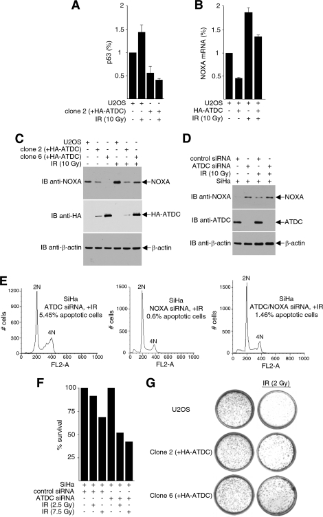
FIG. 5.
ATDC reduces p53 recruitment on the NOXA promoter and represses NOXA expression. (A) Cross-linked proteins from cell extracts were immunoprecipitated with anti-p53 antibodies, and immunoprecipitated DNA were purified and subjected to real-time PCR analysis to detect p53 association with the NOXA promoter. Values were obtained by the Pfaffl method and are presented relative to the input before immunoprecipitation. The data are from one experiment representative of two independent experiments with similar results (error bars, SD of triplicates). (B) Real-time RT-PCR analysis of NOXA mRNA in U2OS cells transfected with HA-ATDC expression plasmids or vectors, with or without IR treatment. The data are representative of three independent experiments with similar results (error bars, SD). (C) Extracts prepared from U2OS cells transfected with the pcDNA3.1HA vector or stably expressing HA-ATDC were subjected to Western blot analysis to detect NOXA expression. (D) Extracts prepared from SiHa cells expressing ATDC siRNA or control siRNA were immunoblotted with the indicated antibodies to assess endogenous NOXA protein expression. (E) SiHa cells transfected with ATDC siRNA, NOXA siRNA, or ATDC siRNA plus NOXA siRNA and treated with 2.5 Gy of γIR. After 24 h, the cells were harvested and analyzed by FACS for apoptotic cells. (F) Cell survival assays were performed on SiHa cells transfected with either ATDC siRNA or control sequences, with or without IR treatment. (G) U2OS cells and U2OS cells expressing HA-ATDC (clone 2 and clone 6) were either treated or mock treated with irradiation. Cell survival assays were carried out as described in Materials and Methods.

