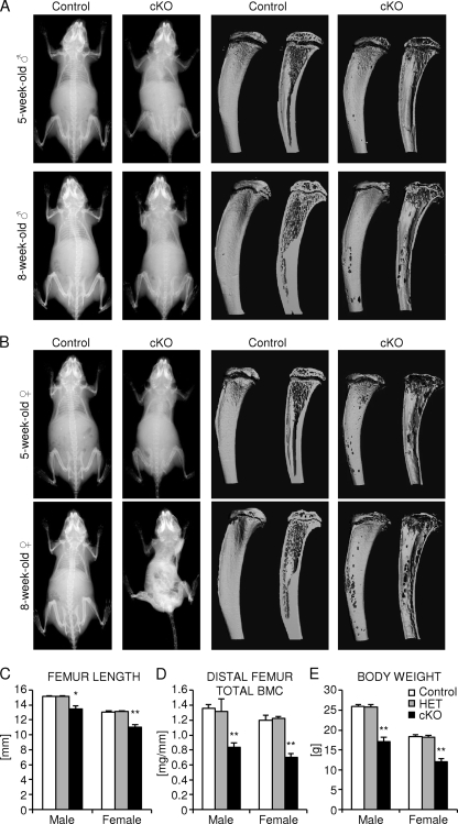
FIG. 3.
Analysis of juvenile osteocyte-specific β-catenin cKO mice. (A and B) Representative whole-body skeletal radiographies (left two columns) and μCT images of the proximal tibia to the midshaft level (right two columns) of 1-month-old (top row) and 2-month-old (bottom row) homozygous osteocyte-specific β-catenin-deficient (cKO) and control littermate male mice (A) and female mice (B). (C to E) Quantification of femoral length (C), cross-sectional total BMC in the distal femur metaphysis (D), and body weight (E) of 2-month-old homozygous (cKO) and heterozygous (HET) osteocyte-specific β-catenin-deficient and control littermates. There were 3 to 11 mice in a group. Values for the mutant that were significantly different from the value for control littermate mice of the same gender using unpaired Student's t tests are shown as follows: *, P < 0.05; **, P < 0.01.

