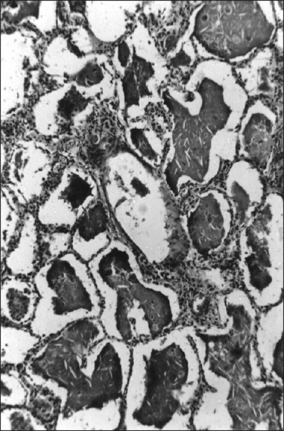
Figure 2.

Histopathological picture of lung section showing alveoli filled with eosinophilic amorphous material; the intra-alveolar material shows cleft-like spaces and foamy macrophages. The lining alveolar cells were swollen. Parenchymal architecture remained intact (H and E, ×50)
