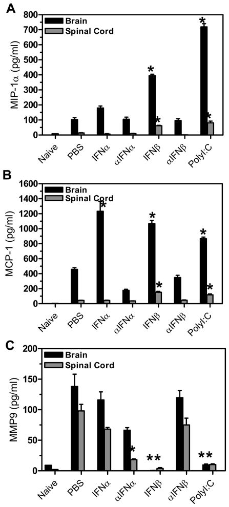
Figure 3. Chemokine and MMP expression was altered in TMEV-infected mice administered IFNβ or polyI:C.
TMEV-infected mice were administered IFNα, αIFNα, IFNβ, αIFNβ, polyI:C or PBS (control) on day 0 and day 4 post infection. The brain and spinal cord was removed from mice (3 mice per group) at 7 days post infection. The RNA was isolated from the organs, converted to cDNA, and real time PCR was conducted with primers specific for MIP-1α (A), MCP-1 (B), or MMP9 (C). The experiment was repeated three times with one representative experiment shown. The stars above the bars represent a significant difference (increase or decrease) in expression compared to control (PBS) treated mice.

