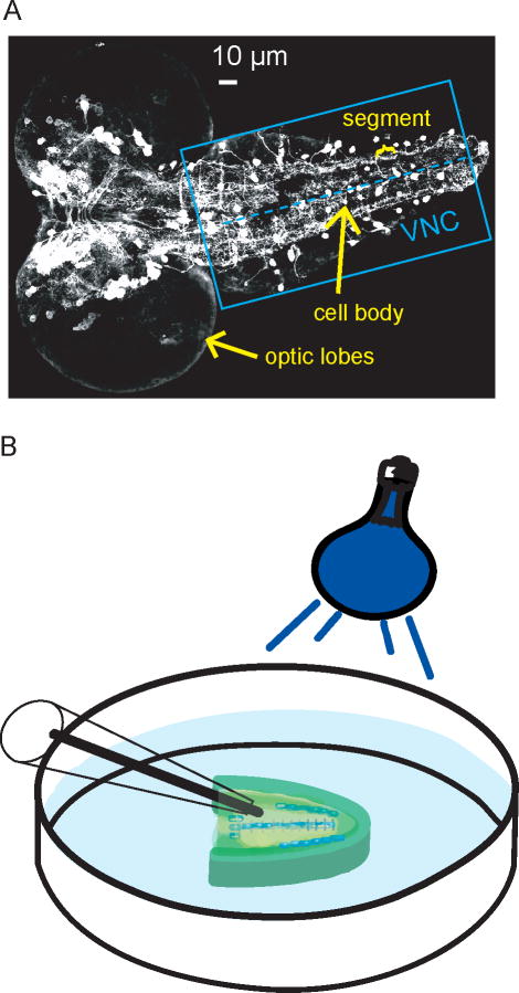
Figure 1.
A) Fluorescence microscopy image of GFP-labeled dopaminergic neurons in a 5-day-old, 3rd instar larva CNS. The blue box indicates the ventral nerve cord, and the dashed line marks the midline, where many cell bodies (white circles) are located. On either side of the midline is the neuropil region, which is rich in dopamine terminals. B) Schematic of microelecrode placement into the neuropil region of the ventral nerve cord with blue-light stimulation. The optic lobes have been removed from the CNS.

