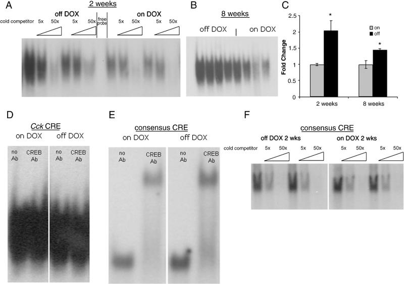
Figure 3.
Protein binding at the Cck promoter. (A, B) Electrophoretic mobility shift assay using the Cck CRE-like site with striatal tissue from animals overexpressing ΔFosB for 2 weeks (A) or 8 weeks (B). In (A), competition with excess unlabeled competitor DNA was performed to demonstrate probe specificity. (C) Binding shown in A and B was quantified using densitometry. (D) Supershift assay using the Cck-like CRE site and a CREB antibody (lanes 2, 4) on lysates from ΔFosB overexpressing mice on (lanes 1-2) or off (lanes 3-4) dox for 2 weeks. (E) Supershift assay using a consensus CRE site and a CREB antibody (lanes 2, 4) on lysates from ΔFosB overexpressing mice on (lanes 1-2) or off (lanes 3-4) dox for 2 weeks. (F) Electrophoretic mobility shift assay using a consensus CRE site with striatal tissue from animals overexpressing ΔFosB for two weeks. Excess unlabeled competitor DNA was used to demonstrate probe specificity. For A, D, E, and F representative experiments are shown. * p<0.05 vs on dox animals.

