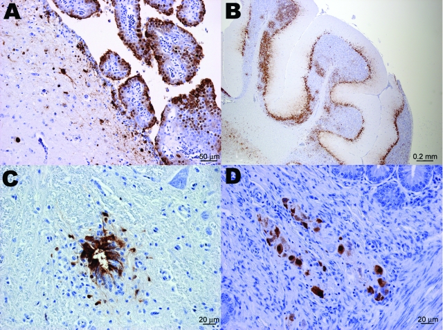
Figure 1.
Immunohistochemical staining for influenza virus nucleoprotein in central and peripheral nervous system of naive juvenile Canada geese tissues after challenge with influenza virus (H5N1). A) Cerebrum. Positive immunolabeling of neurons, glial cells, ependymal and choroid plexus epithelial cells. B) Cerebellum. Extensive positive immunolabeling of Purkinje cells and neurons of the granular layer. C) Spinal cord. Positive immunolabeling of ependymal cells of the central canal and adjacent neurons and glial cells. D) Small intestine. Positive immunolabeling of neurons of the submucosal plexus.

