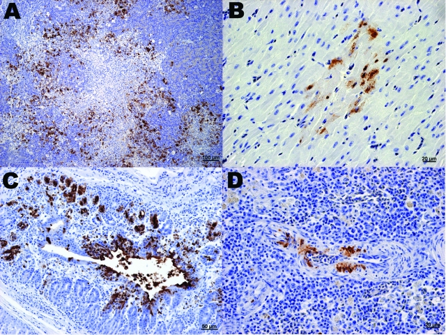
Figure 2.
Immunohistochemical (IHC) staining for influenza virus nucleoprotein in tissues of naïve juvenile Canada geese after challenge with influenza virus (H5N1). A) Pancreas. Large areas of necrosis are surrounded by pancreatic acinar cells with strong positive intranuclear and intracytoplasmic immunolabeling. B) Heart. Positive intranuclear and intracytoplasmic immunolabeling of myocytes. C) Proventriculus. Strong positive immunolabeling of compound tubular gland epithelium. D) Splenic arteriole. Positive IHC staining of vascular smooth muscle cells.

