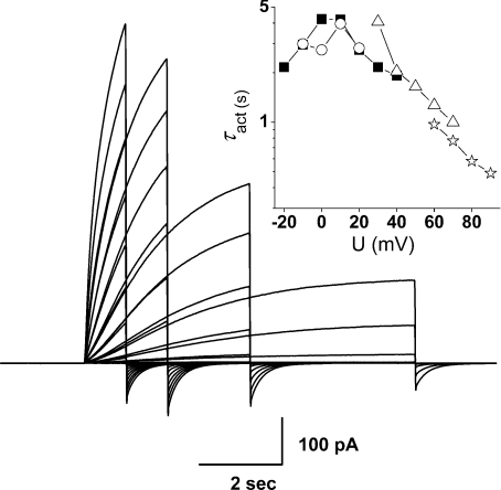
Figure 2. Use of different pulse lengths to evaluate τact at different voltage ranges, to minimize distortion of kinetics by proton depletion.
Superimposed are four pulse families of different durations in the same cell, transfected with the H193A–H140A tandem dimer at pHo 7.0, pHi 6.5 with 10 μm Zn2+. Pulses were applied in increasingly positive steps in 10 mV increments from a holding potential of −60 mV up to +20 mV (8 s pulses, ○), +40 mV (4 s, ▪), +70 mV (2 s, ▵) or +90 mV  . Pulses were applied at 22 s intervals except for a 38 s interval for the 8 s family. The order of families was 4 s, 8 s, 2 s and 1 s. The inset shows τact estimated from these records.

