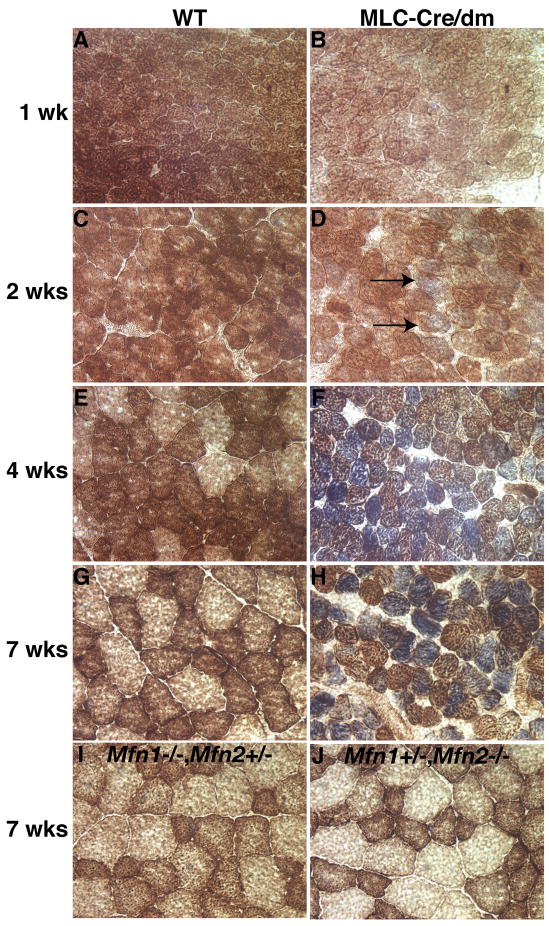
Figure 2. Temporal analysis of respiratory deficiency in MLC-Cre/dm muscle.
Transverse sections of TA muscle were stained histochemically for COX (brown) and SDH (blue) activity. (A, C, E, and G) Wildtype muscle. (B, D, F, and H) Double mutant muscle. Blue staining is indicative of mitochondrial dysfunction. Note the initial appearance of faintly blue fibers in D (arrows) and deeply blue fibers in F and H. The ages of the samples are indicated. (I and J) Genotypes and age of the muscles are indicated. 400× magnification. See also Figure S2.

