Figure 1.
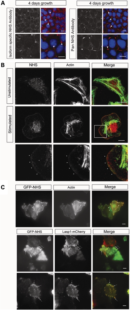
NHS localizes to sites of cell contact, the leading edge of lamellipodia and focal adhesions. (A) Endogenous NHS (red) localized to sites of cell–cell contact in Caco-2 cells, detected by an N-terminal isoform-specific antibody (left panel) and a C-terminal pan NHS antibody (right panel). Lower panel for each antibody staining is a higher magnification. Nuclei were counterstained with DAPI. Staining for NHS (red) was prominent at tricellular contacts (arrowheads). Scale bar 10 µm. (B) Endogenous NHS (detected with pan NHS antibody; red) localized at the leading edge of lamellipodia in stimulated MTLn3 cells at the late 3 min transient (arrowheads). Cells were counterstained for F-actin (green). Top panel, unstimulated; middle panel, 3 min stimulation; bottom panel, enlargement of boxed area showing NHS at the leading edge of the lamellipod. Scale bar 10 µm. (C) Live TIRF imaging of GFP-NHS-1A (green) in MCF7 cells revealed NHS localization at cell junctions (arrowhead), the leading edge and co-localization with actin puncta (arrows) and actin fibres (red, top panel). GFP-NHS-1A co-localized with the focal adhesion complex protein Lasp1 (red, bottom two panels). Scale bar 10 µm.

