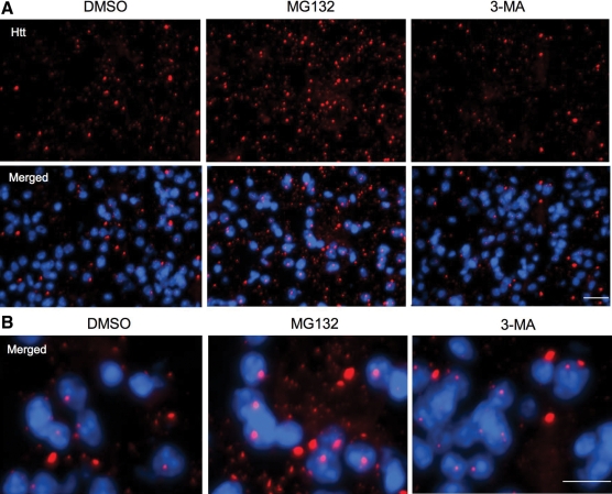
Figure 7.
Immunofluorescent staining of the striatal sections of the 12-month-old HD KI mouse brain. The striatum of HD KI mice was injected with DMSO, the UPS inhibitor MG132, or the autophagy inhibitor 3-MA. After 24 h, the striatal sections were isolated and fixed for immunofluorescent staining with the antibody to htt (mEM48) and Hoechst dye to label nucleus (blue). Note that mutant htt forms nuclear inclusions and neuropil aggregates that are small and outside the nucleus. MG132, but not 3-MA, increased the density of htt aggregates seen in the low (A) and high (B) magnification images. Scale bars: 20 µm in (A) and 10 µm in (B). Two female mice each group were examined.

