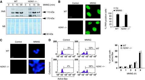
Figure 5.
PARP-1, calpains, and Bax are activated in the absence of H2AX. (A) Poly(ADP-ribose) (PAR) immunoblotting detection in total extracts from WT and H2AX−/− MEFs untreated or treated with MNNG at different times. The membrane was stained with naphtol blue (NB) to assess protein loading. (B) WT and H2AX−/− MEFs were untreated (control) or treated with MNNG (15 min), immunostained for PAR detection, and visualized by fluorescent microscopy. Representative micrographs of each cell type are shown. Data are means±s.d. (n=5). Bar: 10 μm. (C) Fluorescent assessment of calpain activity measured in WT and H2AX−/− MEFs untreated (control) or treated with MNNG (1 h). Representative micrographs of each treatment are shown. This experiment was repeated four times, yielding similar results. Bar: 10 μm. (D) WT and H2AX−/− MEFs were treated with MNNG and Bax activation was measured by flow cytometry. Representative cytofluorometric plots of untreated (control) and MNNG-treated (9 h) cells are shown. Data are the means±s.d. (n=6). A full-colour version of this figure is available at The EMBO Journal Online.

