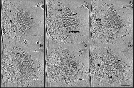
Figure 1.
Cryo-electron tomography of purified human centrosomes. Z slices (20 nm spacing) from a three-dimensional reconstruction (140 slices) of a centrosome, showing longitudinal sections of a centriole and its procentriole surrounded by the pericentriolar material (*: PCM). The procentriole (P) is perpendicular to the proximal part of the mature centriole (C). Arrows point towards microtubule blades. Ring-shaped objects (open arrowheads) are visible in the PCM. The central structure of the cartwheel (ctw) is distinguished in the procentriole (arrowhead in slice 60). The distal extremity of the mature centriole is filled with electron dense material (open arrow in slice 80). Scale bar, 250 nm.

