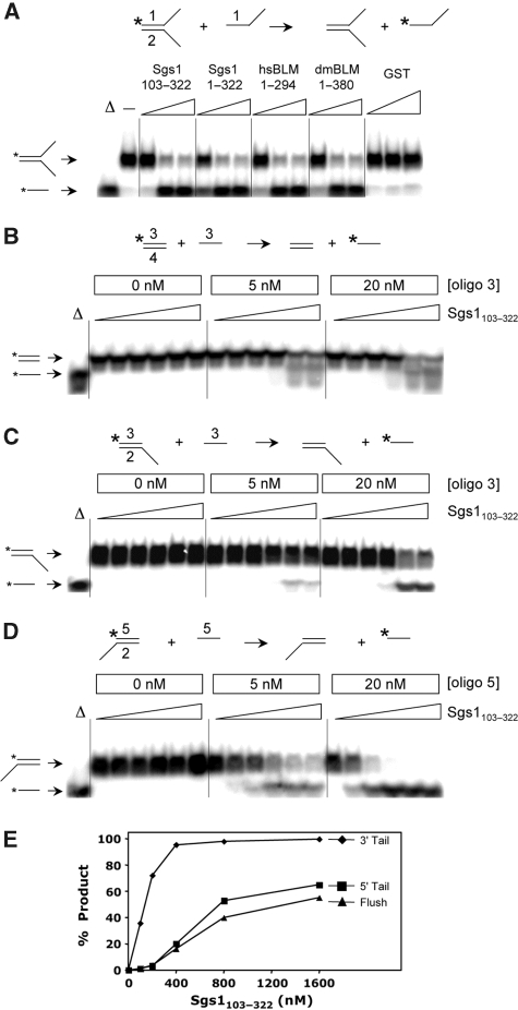
Figure 4.
The SE domains from BLM/Sgs1 orthologs show DNA SE activity. (A) The SE assay is illustrated at the top of the panel. Reactions contained the indicated SE domain proteins at 0 (−), 50, 200 or 400 nM plus 2 nM forked DNA (where oligo #1 is 32P-labelled) plus 10 nM oligo #1. Substrate DNAs were incubated with GST at 50, 200 or 800 nM as negative control. The reactions were incubated at 37°C for 30 min under standard conditions and the products were analysed by 8% PAGE and phosphorimaging. The first lane (Δ) contains 32P-labelled oligo #1 as marker. (B) Sgs1103−322 (0, 100, 200, 400, 800 and 1600 nM) was used in an SE assay using blunt-ended substrate as indicated in the reaction at the top of the panel. Reactions contained 1 nM duplex DNA plus the indicated amounts of oligo 3. Assays were performed as in (A) except for use of 10% PAGE. (C) Sgs1103−322 was assayed using 5′-tail duplex DNA as substrate as in (B). (D) Sgs1103−322 was assayed using 3′-tail duplex DNA as substrate as in (B). (E) The SE reactions shown in (B–D) (20 nM unlabelled oligo) were quantified and are presented as a function of protein concentration. Sequences of the indicated oligos are presented in Supplementary Table 1. Throughout, all proteins are His6-tagged. Asterisks represent positions of 32P-labelling.

