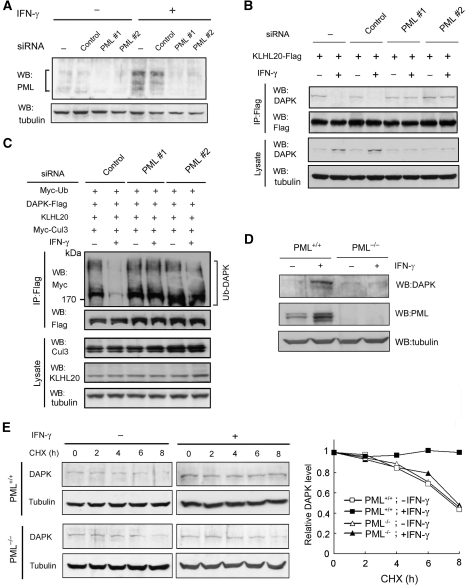
Figure 6.
PML depletion reverses the inhibitory effects of IFN on DAPK ubiquitination and degradation. (A) Generation of PML knockdown cells. HeLa cells were infected with lentivirus carrying indicated siRNA and selected with puromycin. Cells were treated with or without IFN-γ for 18 h and then lysed for western blot analysis. (B) PML siRNA rescues the interaction between DAPK and KLHL20 in IFN-γ-treated cells. Cells as in (A) were transfected with KLHL20-Flag and treated with IFN-γ for 18 h. Cells were lysed for immunoprecipitation with anti-Flag antibody. The immunoprecipitates and cell lysates were analysed by western blot with antibodies as indicated. (C) PML siRNA rescues KLHL20-mediated DAPK ubiquitination in IFN-γ-treated cells. Cells as in (A) were transfected with indicated constructs and treated with IFN-γ for 18 h. DAPK ubiquitination was analysed as in Figure 2A. The expression levels of various proteins are shown on the bottom. (D) IFN-γ fails to upregulate DAPK in PML null cells. PML+/+ or PML−/− MEFs were treated with IFN-γ for 18 h and then lysed for western blot analysis with indicated antibodies. (E) PML is required for IFN-γ-induced DAPK stabilization. Cells as in (D) were treated with IFN-γ for 18 h and then with 50 μg/ml cycloheximide for indicated time points. Cells were lysed for western blot analysis and the level of DAPK was normalized to that of tubulin and plotted on the right.

